La vidéo de présentation est disponible à ce lien



Communauté de la médecine nucléaire
Partagez vos présentations ppt, pdf, video…
La vidéo de présentation est disponible à ce lien
Carcinoses péritonéales et imagerie TEP-TDM
Bases physiopathologiques et zones forteresses
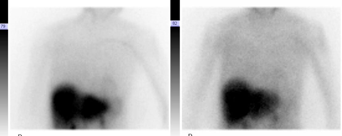
Présentation de 2010 (MednucLR) mais qui peut encore servir si votre neurologue préféré vous demande une MIBG cardiaque ^^
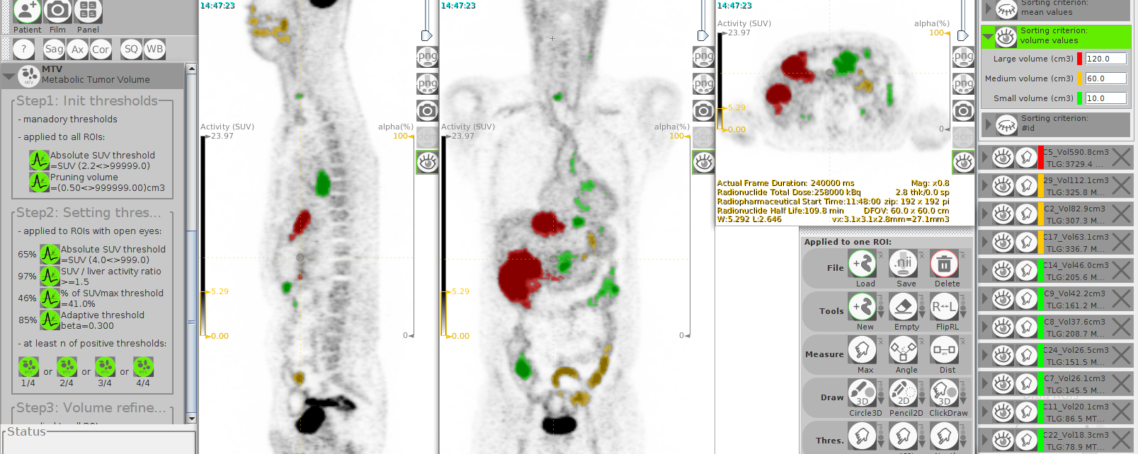
Site internet : http://www.lifexsoft.org
Texture analysis is gaining considerable interest in medical imaging, in particular to identify parameters that might characterize tumor heterogeneity. We developed an easy-to-use freeware enabling calculation of a broad range of conventional, textural and shape indices from PET, MR and CT images.
The software is written in Java and does not rely on any commercial libraries. LIFEx is dedicated to researchers, radiologists, nuclear medicine physicians and oncologists and includes two operation modes:
Read more about Available levels.
LIFEx reads DICOM images locally or over a network using a DICOM browser, is compatible with Osirix and includes a powerful 3D reconstruction-based slice viewer. Volumes of interest (VOI) can be either imported from previously created files or drawn and manipulated using LIFEx. Results are exported in Excel format files. LIFEx runs on Windows, MacOs and Linux. It is distributed with examples and includes a tutorial. User support is available for the “validated” mode. Users can optionally contribute to the gathering of index values measured in different tissue types and different images as a public data bank of reference values is currently being built and integrated in the software for assisting the users with the interpretation of their results.
LIFEx has already been distributed to research labs, nuclear medicine departments and radiology departments for investigating different tumor types (gliomas, cervix, lung, breast, and colorectal tumors), and has been very positively received. The intuitive interface associated with the tutorial made it fast to master for staff, and allowed us to start building databases of normal textural values in brain (white and grey matter), breast, liver, lung, fat, and muscles for various imaging equipment and protocols in PET and CT, while MR data are currently being collected and processed. Such data enable an accurate characterisation of the variability of different textural indices in a given imaging modality as a function of the scanner and the imaging protocol. New indices are being implemented based on users requests as the software is intended to evolve over time based on advances in the field.
3èmes JFMN – Nantes
Atelier parrainé GE – Vendredi 19 mai 2017
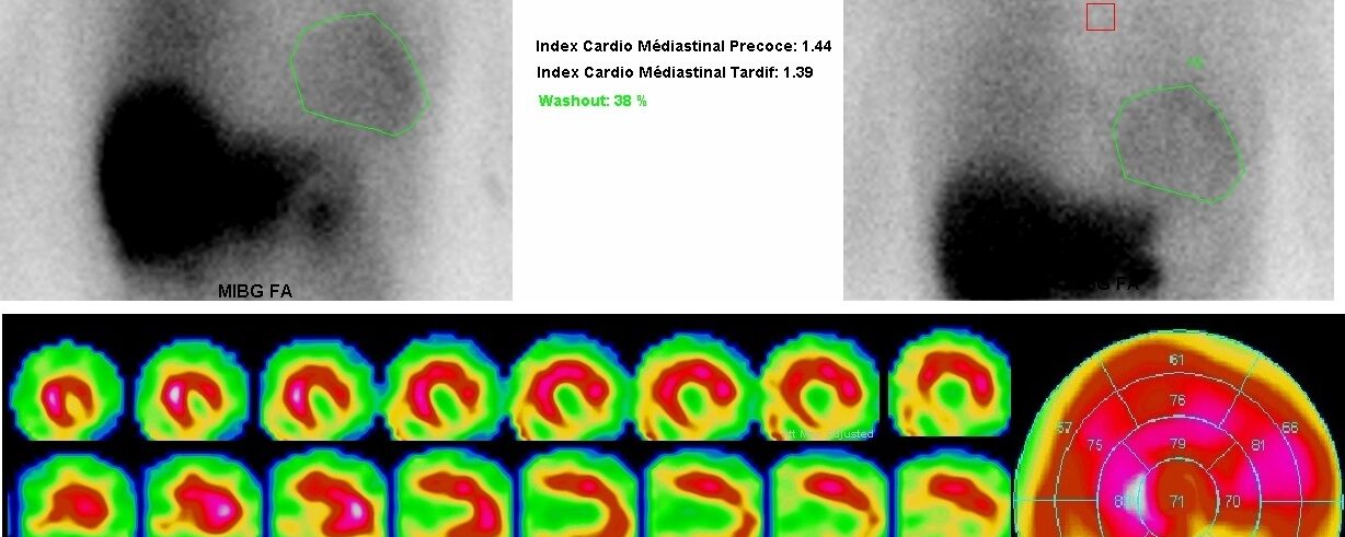
Présentation réalisée pour la réunion Mednuc-LR de septembre 2016,à partir d’un cas clinique Nîmois.
Ce document fait la synthèse d’un document de la SoFRA sur les extravasation de Médicaments Radiopharmaceutiques utilisés en diagnostic et thérapie.
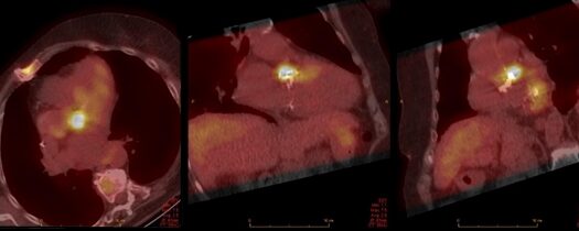
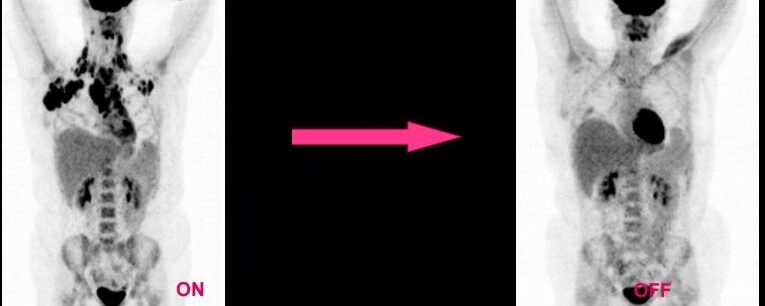
Présentation sur la TEP au FDG dans l’évaluation thérapeutique des lymphomes,réalisée pour le service de MN de Perpignan.
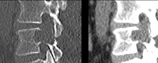
Tumeurs osseuses 2/2: bénin
Cas cliniques, diapos de synthèse et arbre décisionnel
Tumeurs osseuses 1/2: malin
Cas cliniques, diapos de synthèse et arbre décisionnel
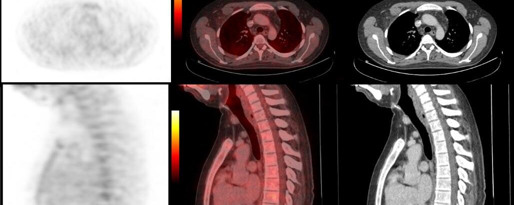
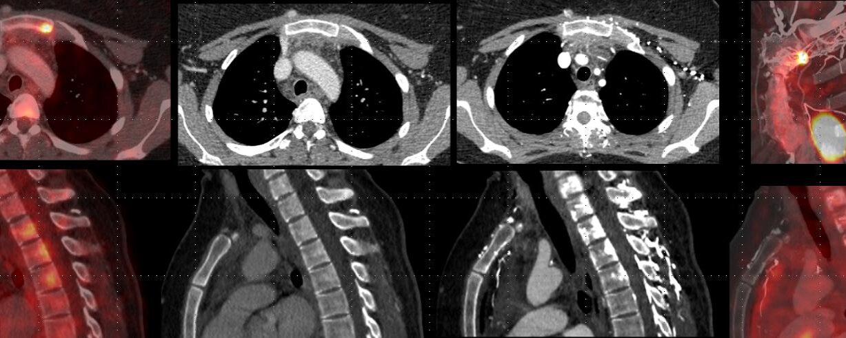
Présentation sur les lésions tumorales du médiastin antérieur en TEP 18F-FDG